The sensation of persistent ear pressure, often described as a feeling of fullness, dull pain, or being underwater, is one of the most common, yet frustratingly elusive, complaints in Otolaryngology. While many associate it with simple altitude changes or a common cold, chronic or recurring ear pressure frequently points to an underlying malfunction of the Eustachian Tube (ET). This small, membranous, and cartilaginous canal connects the middle ear to the back of the nasopharynx, and its singular, non-negotiable role is to equalize the air pressure between the middle ear space and the external atmosphere, as well as to drain any normal middle ear secretions. When this tube fails to open or close properly—a condition termed Eustachian Tube Dysfunction (ETD)—the resulting pressure imbalance and negative pressure within the middle ear can lead to a host of debilitating symptoms, including muffled hearing, crackling or popping noises, and chronic discomfort. Diagnosing ETD requires more than a simple visual inspection of the eardrum; it necessitates a sophisticated, multi-modal assessment that combines detailed clinical history, advanced objective testing, and the critical differentiation between ETD and other conditions that mimic its symptoms, such as Temporomandibular Joint (TMJ) disorder or even subtle inner ear issues.
The Most Common, Yet Frustratingly Elusive, Complaints in Otolaryngology
Persistent ear pressure presents a significant diagnostic challenge precisely because the primary symptom is subjective and non-specific. The most common, yet frustratingly elusive, complaints in Otolaryngology can stem from mechanical, inflammatory, or neurological issues, all presenting with the feeling of fullness. Patients often report difficulty hearing their own voice (autophony) or a sense that their ears need to “pop,” providing clues that the equalization mechanism is flawed. The true difficulty for the clinician lies in confirming the functional impairment of the Eustachian Tube. Since the ET is a deep, internal structure, direct visualization is impossible, forcing practitioners to rely on indirect measurements that infer the tube’s functional status by monitoring its effects on the pressure within the middle ear. A comprehensive diagnosis starts not with a test, but with an exhaustive interrogation of the patient’s symptom triggers, duration, and patterns of fluctuation.
The Singular, Non-Negotiable Role Is to Equalize the Air Pressure
Understanding the mechanics of the ET is key to diagnosing its failure. The singular, non-negotiable role is to equalize the air pressure between the sealed middle ear cavity and the outside world. Under normal circumstances, the ET remains closed, only opening briefly during swallowing, yawning, or chewing. This brief opening allows a tiny bubble of air to enter the middle ear, neutralizing any negative pressure that may have developed due to the natural absorption of air by the middle ear lining. ETD typically manifests as a failure of this crucial opening mechanism—a “failure to ventilate.” When the tube remains functionally blocked, the air inside the middle ear is slowly absorbed, creating a sustained negative pressure that pulls the eardrum inward, resulting in the feeling of fullness and muffled sound transmission.
The Standard First-Line Objective Test Is Tympanometry
The most rapid and accessible objective tool for assessing middle ear pressure is a simple, non-invasive procedure. The standard first-line objective test is Tympanometry, which measures the mobility of the eardrum (tympanic membrane) as air pressure is varied in the external ear canal. In a healthy ear with a patent ET, the eardrum is most mobile when the pressure in the external canal is equal to the ambient pressure of the middle ear (a flat tracing). A characteristic finding in ETD is a negative middle ear pressure tracing, indicating that the middle ear pressure is significantly lower than the outside pressure, confirming the presence of a vacuum-like effect that pulls the eardrum inward and restricts its movement, visually confirming the physiological consequence of a blocked ET.
ETD Requires Sophisticated, Multi-Modal Assessment
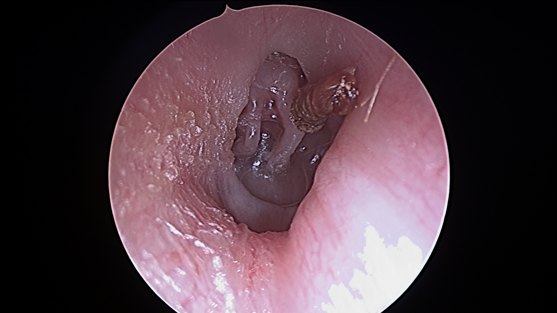
Because tympanometry alone cannot definitively determine the cause of the ET failure, the diagnostic process must be escalated. ETD requires sophisticated, multi-modal assessment that often involves specialized functional tests. The Eustachian Tube Function Test (or patency testing), often conducted in conjunction with tympanometry, attempts to quantify the tube’s ability to equalize pressure in response to deliberate pressure changes. Furthermore, a nasal endoscopy may be necessary to directly inspect the pharyngeal opening of the ET—known as the Torus Tubarius—to check for anatomical obstructions such as large adenoids, nasal polyps, or mass lesions, particularly in persistent, unilateral (one-sided) cases where a tumor is a rare but critical differential diagnosis.
Imaging Is Usually Not Warranted Unless a Mass Lesion Is Suspected
For routine, bilateral ETD following an upper respiratory tract infection, advanced imaging holds little diagnostic value. Imaging is usually not warranted unless a mass lesion is suspected or if the patient is unresponsive to standard therapy. In cases of chronic, treatment-resistant ETD or unilateral fluid accumulation (effusion), a high-resolution CT scan or MRI may be ordered. This imaging is not used to visualize the ET itself, but rather to assess the surrounding anatomy—specifically to check for tumors in the nasopharynx that could be physically compressing or blocking the tube’s opening, or to evaluate the extent of inflammatory changes in the mastoid bone that might be contributing to chronic middle ear fluid.
Other Conditions That Mimic Its Symptoms Must Be Carefully Ruled Out
A major hurdle in diagnosis is the high overlap in symptoms with entirely non-otologic conditions. Other conditions that mimic its symptoms must be carefully ruled out before settling on an ETD diagnosis. Temporomandibular Joint (TMJ) disorder, characterized by jaw pain, clicking, and limited movement, is a frequent mimicker. The nerves and muscles that control the jaw are anatomically close to the middle ear and the ET, and chronic tension or dysfunction in the TMJ area can cause referred pain and a sensation of ear fullness that is easily confused with ETD. Similarly, Cervical Spine issues, Sinusitis, and certain forms of Vestibular Migraine can all present with ear pressure, demanding a truly differential diagnostic approach that integrates ENT, dental, and neurological assessments.
Patulous ETD Is Characterized by the Tube Remaining Open Most of the Time
While the majority of ETD cases involve a tube that remains stubbornly closed (obstructive ETD), a less common but highly distinctive form exists where the tube remains abnormally open. Patulous ETD is characterized by the tube remaining open most of the time, leading to a different, specific set of symptoms. The most classic symptom is autophony, where the patient hears their own voice, breathing, and even heartbeats intensely echoing within their head because the open ET transmits these sounds directly from the nasopharynx to the middle ear. Unlike obstructive ETD, which often benefits from decongestants, PETD can be worsened by dehydration or exercise. Diagnosis is often heavily reliant on the highly specific patient history, as objective tests can sometimes appear normal.
The Most Common Underlying Cause of Eustachian Tube Dysfunction is Inflammation
For the average patient with transient ETD, the problem is usually rooted in inflammation that swells the delicate lining of the tube. The most common underlying cause of Eustachian Tube Dysfunction is inflammation from an upper respiratory tract infection (the common cold), allergic rhinitis, or chronic sinus issues. This mucosal swelling narrows the tube’s lumen, preventing air passage. The initial therapeutic approach in these cases is conservative, focusing on reducing this inflammation with nasal steroid sprays, oral decongestants (used cautiously and briefly), and antihistamines, all aimed at shrinking the swollen tissues around the ET opening and allowing it to vent naturally. Only when these medical treatments fail to resolve the pressure does the focus shift to procedural intervention.
Procedural Interventions Aim to Physically Force the Tube Open
When chronic ETD fails to respond to months of aggressive medical management, the possibility of permanent or long-term procedural intervention is explored. Procedural interventions aim to physically force the tube open or bypass its function altogether. A long-standing solution for ventilating the middle ear is the placement of tympanostomy tubes (pressure equalization tubes) through the eardrum, which temporarily takes over the ET’s function of pressure equalization. A newer, minimally invasive technique is Balloon Dilation of the Eustachian Tube (BET), where a tiny balloon catheter is inserted into the ET and briefly inflated to remodel the cartilage and forcibly widen the passageway, aiming for a permanent restoration of the tube’s natural function.
A Successful Diagnosis Is Ultimately a Triangulation of Patient Experience and Objective Data
Ultimately, navigating the diagnostic maze of ear pressure requires the clinician to act as a detective, synthesizing varied and sometimes contradictory information. A successful diagnosis is ultimately a triangulation of patient experience and objective data, recognizing that no single test provides the final answer. The practitioner must weigh the patient’s subjective report of popping and fullness against the objective tympanometry reading, the endoscopic visualization, and the response to initial medical management. This synthesis allows the specialist to confidently confirm ETD, rule out the less common but critical differentials like masses, and embark on a targeted treatment plan designed to restore the normal physiological rhythm of the middle ear—a true return to pressure equilibrium.